Multislice Computed Tomography
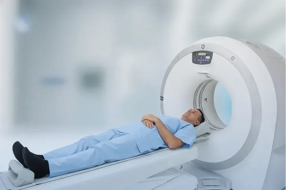
In computed tomography (CT), there is an X-ray tube that emits X-rays and detectors that capture these rays. The patient lies between these two components. The X-rays pass through the patient's body and are then detected by the detectors (see Image 1). Since the atomic densities of the body's tissues vary, each tissue absorbs a different amount of X-rays, which creates the tissue images. The tube and detectors rotate 360 degrees around the patient to capture cross-sectional images of the examined area.
In conventional tomography, after an image of one slice is taken, the table moves forward slightly to capture the next slice. This method is both time-consuming and highly sensitive to respiratory movements, making it unsuitable for cardiac and vascular examinations.