Ultrasonography
With our state-of-the-art devices, almost all vascular diseases can be investigated, and all ultrasonography examinations can be performed in color.
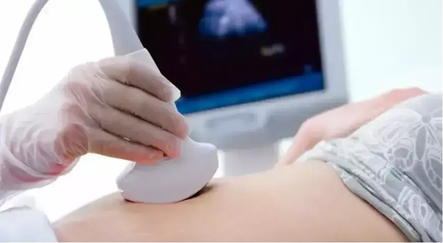
Ultrasonography is an imaging technique that uses ultrasound waves. It is widely used for preliminary diagnosis of many diseases, especially effective for examining organs like those in the abdomen where sound waves can easily pass through. This method does not use X-rays. Ultrasound is a diagnostic technique that images internal organs by using sound waves at frequencies too high for the human ear to hear. Ultrasound does not involve radiation, making it safe for use in pregnant women and infants. The sound waves emitted from the device are reflected back from the patient’s body and detected by the same device. The differences in reflections vary between organs. Therefore, structures with different textures produce different images. Tumors or cysts within normal tissues reflect sound waves differently, appearing distinct and allowing diagnosis. During imaging, the “probe” is moved over the patient’s body, and cross-sectional images of the underlying area, as well as moving organs, appear on the screen. The radiologist makes the diagnosis during this process. The images obtained have limited contribution to the diagnosis. Ultrasound procedures are performed by radiologists who have received specialized ultrasound training during their residency.