Artificial Intelligence and Mammography
Today, mammography is the primary screening method for breast cancer, the most common cancer among women. Breast MRI and tomosynthesis are also frequently used in breast cancer screening. Due to the high dimensionality and complexity of images obtained from these imaging methods, evaluating their content is challenging and time-consuming. To increase radiologists' efficiency and accuracy in reading breast images, many computer-aided software programs have been developed.
Radiological images are not just pictures; fundamentally, they are digital data in matrix format. With the emergence of machine learning and deep learning algorithms, revolutionary advances have been witnessed. Since 2012, research on this topic has increased exponentially due to the success of deep learning algorithms in image analysis. The achievements of deep learning methods and convolutional neural network algorithms in other image analysis fields have led to their use in medical image analysis and mammography reading.
One advantage of deep learning is that the system can learn by itself; instead of humans defining image features and teaching the computer the relevant calculations, deep learning allows computers to learn image features on their own. In other words, the stage has shifted from humans teaching image features to computers independently learning those features.
Computer-Aided Detection (CAD) software was first developed in the early 1990s for breast cancer detection in mammography.
CAD systems are important for minimizing missed or misinterpreted lesions in mammography. New generation deep learning-based CAD systems help improve breast cancer screening programs. Studies have shown that using AI-supported CAD as a decision support tool helps radiologists more than traditional approaches. Moreover, research shows that breast radiologists assisted by AI-enhanced decision support systems have higher diagnostic performance compared to reading alone.
Artificial intelligence is definitely influencing radiology faster than other medical fields. In just a few recent years, various applications have been developed that reach or even surpass human performance in specific image recognition tasks like breast imaging. The next step is the integration of these applications into daily practice, following necessary approvals and legal frameworks.
In the future, deep learning-based AI systems will increase efficiency and confidence in the daily routine of breast radiologists, allowing them to devote more time to the patient's clinical care. Radiologists will shift their focus from the images themselves to the clinical context, aided by AI-powered decision support systems.
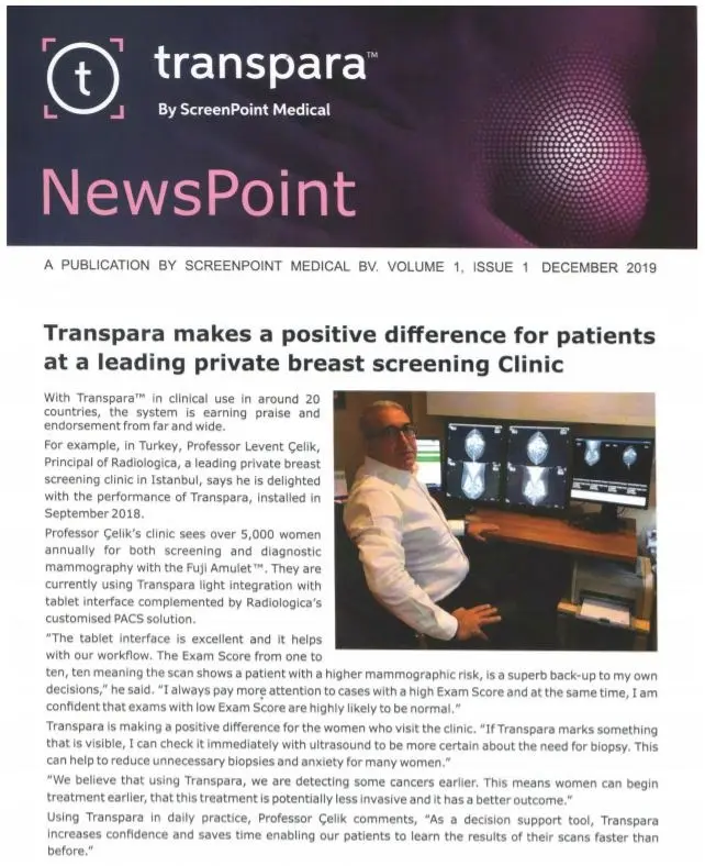
Since October 2018, our clinic has been using the AI system Transpara by Screenpoint Medical, which received FDA approval in 2019. We have contributed to its development by providing labeled data, and currently employ it in mammographic breast cancer screening.
Transpara by Screenpoint Medical