What is Contrast-Enhanced Mammography?
When and Why is it performed?
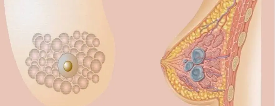
Because breast cancers have a high degree of vascularization, they show prominent enhancement compared to normal breast tissue when contrast is administered. This principle forms the basis for more detailed evaluation of breast cancer with breast MRI examinations.
With special technical equipment and software, it is also possible to visualize the uptake (tumor enhancement) of iodinated contrast agent in mammography. Mammography examinations performed by injecting contrast agent intravenously are called contrast-enhanced mammography (spectral mammography, dual-energy contrast mammography).
In Dual-Energy Contrast Mammography (Spectral Mammography), the physical principle is based on the different absorption of X-rays at high and low energies by the iodinated contrast agent.
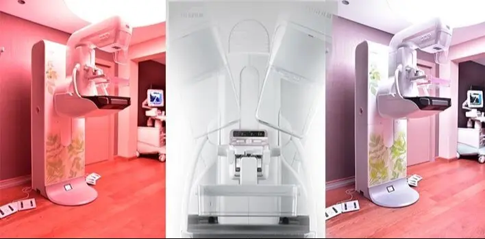
During contrast-enhanced mammography, iodinated contrast agent (the same as used in CT scans) is administered intravenously, followed by imaging with mammography devices equipped with the necessary hardware and software.
During a single imaging session, the digital mammography device performs both a normal mammogram with low-energy X-rays and a high-energy X-ray exposure (45-50 kV). The low-energy images are also used as standard digital mammography images.
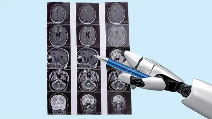
Using developed software, the computer combines the high- and low-energy images to generate a diagnostic image that highlights only the areas of contrast uptake. These images clearly demonstrate tumors with increased vascularization in the breast and allow detection of tumors hidden within dense fibrocystic breast tissue.